 Protocol
AUTHORS
Louise Comas, Bethany Carlisle, Angie Patterson
OVERVIEW
In order to view and quantify symbiotic fungal association in roots, contents of root cells need to be cleared, then fungal walls need to be stained to show arbuscular mycorrhizal (AM) fungal structures. The percentage of root length that has AM colonization can then be assessed.
BACKGROUND
This protocol has been optimized for delicate fine root tissues of woody plants and herbaceous plants. We often work from dried tissues because we are interested in quantifying AM colonization on the same samples that we have assessed root morphological measures such as specific root length (SRL), which requires drying root tissue to obtain its dry mass. Newly-produced fine roots, especially from some species, and when previously dried, are particularly delicate and require tender handling as outlined here.
MATERIALS/EQUIPMENT
- 10% w/v KOH
- ammonia/hydrogen peroxide solution (recipe: 3ml 20% NH2OH in 30ml 3% H2O2Grace C, Stribley DP 1991 Myc Res 95:1160-1162, note both are over-the-counter strengths)
- 5% HCL (10mL v/v glacial HCl in 190mL dH20)
- 0.05% Tryphan blue stain (10 mL glacial acetic acid, 200mL glycerol, 0.2g trypan blue, 190mL dH20, *to accelerate, warm the solution)
- lactoglycerol (1:1:1 lactic acid, glycerol, and water)
- 20 ml glass scintillation vials
- tea strainers
- tweezers
- slides & cover slips (50x75x1 mm slides and 48×65 mm No 1 cover slips)
- mounting medium – typically Polyvinyl-Lacto-Glycerol (PVLG) (In Treseder lab protocols: https://webfiles.nacs.uci.edu/treseder/public/Protocols/PVLG Slide Mounting Medium.pdf)
- compound microscope with a 20x optical lens, a movable stage, and one eye piece fit with either a micrometer or crosshairs
- counter with 4 parts (or combination of a couple of counters to total 4 parts)
PROCEDURE
Clearing and staining
- Clearing step: Place dried roots in a 20 ml glass scintillation vial with enough 10% w/v KOH to cover. Allow roots to soak at room temperature for 1-4 days (~24-48 hrs for herbs, approximately 48-96 hours for woody plants).
- Check samples under the scope to see if stele can be seen through the cortex. If cell contents don’t look cleared, leave to soak in KOH one more day then check again. When cell contents of roots are cleared, pour out KOH using the neck of the scintillation vials to catch roots, pouring KOH through a tea strainer to catch any roots that might escape. As roots become soft from processing, this saves handling them.
- Leaching pigment: (optimize this step as needed for some roots of woody species – roots of woody species that have pigments might only need to be bleached following KOH but EtOH may assist in removing excessive pigments: Dark pigmentation may be removed by soaking in 85% ethanol – samples can be left in EtOH if a break is needed.) Add 85% EtOH to cover following removal of KOH. Multiple exchanges of EtOH may be effective. Move on to step 4 once no more pigment leaches out.
- Bleaching step: (typically not necessary for herbaceous plants) Remove ethanol or KOH and add ammonia/hydrogen peroxide solution to cover roots (recipe: 3ml 20% NH2OH in 30ml 3% H2O2Grace C, Stribley DP 1991 Myc Res 95:1160-1162, note both are over-the-counter strengths). Soak at room temperature for 15 min. Alternatively 3% or 30% H2O2could be used depending on pigmentation of samples. Note: extreme caution should be used because too much H2O2can reduce the ability of fungal cell walls to stain with Tryphan blue.
- Rinse in water and view under the dissecting scope to determine if samples are clear/free enough of pigments. If yes, move to next step. If not, give another day in KOH at room temperature or 5 minutes of clearing in KOH at 65∘C (if cell contents appear viscous or stele isn’t visible), or 10 min of bleaching at room temperature (if cells look clear but pigmentation is still too heavy). (NOTE: extreme caution should be used if herbaceous species and some woody species with particularly tender roots, e.g. Liriodendron, Fraxinus and Sassafras, are cleared in 10% KOH with heat because they will fall apart; for these species, if more clearing time is needed either soak at room temperature or use 1% KOH with heat)
- Preparation for staining: Soak samples in 5% HCL (10mL v/v glacial HCl in 190mL dH20) for 5 minutes at room temperature.
- Staining step: Remove HCl and add enough 0.05% Tryphan blue stain to cover samples. Place in hot water bath of 65∘C for 20 min. Alternatively, roots can be soaked at room temperature for 1-4 hrs as needed by species and strength of stain. Careful not to over-stain because de-staining is not easy. Stain can be saved and reused several times until it starts looking less dark.
- Rinse three times with water and check under the microscope at same magnification that they will be scored. If roots are well stained without being too dark, place in lactoglycerol (1:1:1 lactic acid, glycerol, and water). If mycorrhizas aren’t well stained, return to staining step, checking every 5 minutes. If tryphan blue is too dark, de-stain either in water if just slightly too dark or 50% glycerol if very dark for as long as needed.
Mounting roots on slides
- Add a few drops of mounting medium to the slide.
- Stretch out stained roots in rows across the long dimension of the slide taking care to avoid having roots cross. (NOTE: root length is not determined from these samples, using line-intercept methods to estimate root length requires roots to be randomly distributed)
- Cover with cover slip. Wipe edges with EtOH and let slides cure on a tray lined with paper towels in a drying oven set to 60∘C for 2 days or until set.
Quantifying AM colonization
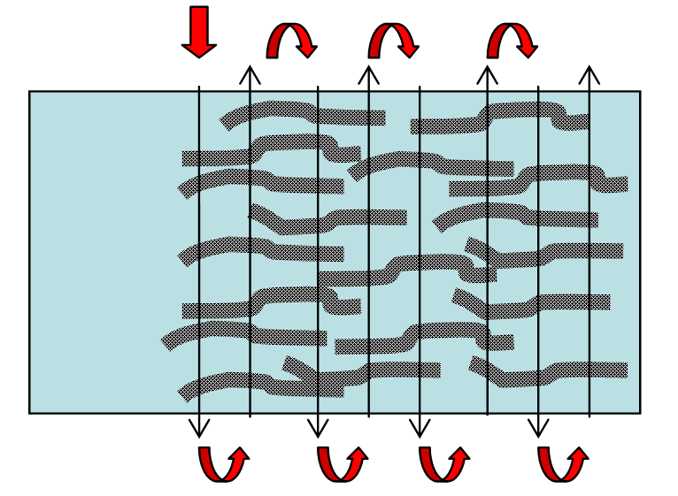
- Count 50-100 root interceptions per sample. 50 intercepts is the minimum with each intercept determining 2% of the total; 100 intercepts is preferred.
- Get a rough count of how many rows of roots have been laid across the slide to determine about how many passes through the slide will be made so that you can space the passes roughly equally distributed across the slide.
- Position the slide to start counts of root intercepts at one end. Rotate the micrometer or crosshairs to give you a vertical line in the field of view.
- Move the stage of the microscope scoring roots for colonization at each intersection point with the vertical line as follows:

- At each point where a root intersects the vertical line, examine the entire cross section of root perpendicular across the root at the point where the root intersects the vertical line, eye-balling the cross section that is 90° perpendicular to the root:

- Focus up and down to check for mycorrhizal structures in all planes of the root cross section, paying attention not to score hyphae that might just be on the surface.
- At each root intersection, score only one of the following four categories: no mycorrhizal structures, arbuscules, vesicles, or mycorrhizal hyphae.
- If arbuscules and vesicles or hyphae are present, mark only arbuscules.
- If vesicles and hyphae are present, mark only vesicles.
- A mark for hyphae should indicate that hyphae alone were seen.Mycorrhizal hyphae should clearly be within the root and not be dark septate fungi. Mycorrhizal hyphae inside roots are typically thicker than those exterior and have uneven/bumpy edges. Non-mycorrhizal hyphae are often thinner with smooth sides. Hyphae of dark septate fungi are often finer than mycorrhizal hyphae and are medium to dark brown with regularly spaced septate sections. Hyphal coils in the cells should be marked as arbuscules. Mycorrhizal vesicles can be especially tricky to learn to identify. Saprotrophic vesicles are often smaller than mycorrhizal vesicles and can sometimes occur more than one to a cell. Good information on the identification of fungal structures can be found online
http://mycorrhizas.info/vam.html
The total of all four categories should equal the total number of root intersections scored. Mycorrhizal colonization is assessed as the percent of the total root intersections that have mycorrhizal fungi present in the root (e.g. the percent of root length that is colonized with arbuscules, vesicles or mycorrhizal hyphae).
- Example: 100 intersections = 15 arbuscules + 3 vesicles + 7 mycorrhizal hyphae + 75 no mycorrhizal structuresTotal colonization = (15+3+7)/(100) = 25% colonization

NOTES AND TROUBLESHOOTING TIPS
Neutral red has sometimes been recommended for staining roots digitized for morphological measures to insure good contrast if automated settings are not available. EtOH can remove some of this stain so that mycorrhizal colonization can be assessed but does not remove it completely so this material is difficult to assess for colonization. To insure high quality scans of the material, software that has automated settings for brightness and contrast can be used in lieu of staining the roots for contrast. If you are working with particularly delicate roots, 1% KOH may also be used for clearing with or without heat. PROTOCOL IS USEFUL FOR – Fresh fine roots from field: e.g. tomato; Dried fine roots from greenhouse and field: e.g. Acer, Populus, Cornus, Nyssa, Liriodendron, Fraxinus.
LINKS TO RESOURCES AND SUPPLIERS
Treseder lab protocols: https://webfiles.nacs.uci.edu/treseder/public/Protocols/PVLG Slide Mounting Medium.pdf
Identification of fungal structures: http://mycorrhizas.info/vam.html
LITERATURE REFERENCES
Mcgonigle TP, Miller MH, Evans DG, Fairchild GL, Swan JA. 1990. A new method which gives an objective-measure of colonization of roots by vesicular arbuscular mycorrhizal fungi. New Phytologist 115(3): 495-501.
Grace C, Stribley DP 1991. Myc Res 95:1160-1162. (bleach recipe) Koske RE and Tessier B, 1983. Mycol. Soc. Am. Newsl. 34(2):59. (PVLG recipe)
HEALTH, SAFETY & HAZARDOUS WASTE DISPOSAL CONSIDERATIONS
Protective measures should be used to prevent contact with Tryphan blue. All liquid with Tryphan blue or trace amounts of it will need special hazardous waste disposal. Solid waste containing Tryphan blue will also need hazardous waste disposal.