 Protocol
AUTHORs
Rosemary White, Ulrike Mathesius
OVERVIEW
This protocol is for making root cross-sections using a vibrating microtome, and can be used for other small tissues. It is very useful for serial sectioning soft tissues, as the agar/agarose supports the tissue which can become compressed using other methods.
BACKGROUND
This is one of a series of protocols on sectioning unembedded plant tissues, prepared by Rosemary White.
Linked protocols
Making hand sections without support material
Using other plant tissues as support tissue to make hand sections
Using support tissue in a hand microtome to make hand sections
Sliding or sledge microtome sectioning of fresh or fixed tissues
Cryostat sectioning of frozen tissues
MATERIALS/EQUIPMENT
- Agarose – ~3% is good for most roots1-2% for very soft roots
- >3% for harder roots and other tissues
- regular Progen DNA grade agarose, gels at 35-37∘C, melts at >90∘C, is OK
- embedding moulds, either commercial or homemade (homemade illustrated below)
- water bath, set at about 60∘C, depending on % agarose used
- cyanoacrylate-based superglue
- Sharp razor blades – single-edged for trimming agarose blocks before glueing downdouble-edged carbon steel for sectioningsapphire blades are great, but expensive
- piece of pink dental wax to trim agarose blocks on
- water – conical flask with plastic pipette, not a wash bottle
- reasonably fine forceps
- fine paintbrush and/or sharpened orange sticks to transfer sections
- sticky slides e.g. poly-L-lysine-coated slides
- Petri dish with moist filter paper to keep sticky slides while collecting sections
UNITS, TERMS, DEFINITIONS
PROCEDURE

- Melt agarose in microwave (remember to loosen lid first) then place agarose bottle in the waterbath to keep it liquid. Also place a small flask of water in the bath, and put some plastic pipettes in this – so you can withdraw liquid agarose from the bottle without it solidifying and clagging up the pipette. Place the embedding mould on a support in the bath so it’s just above the water surface – to keep it warm.
- Fill embedding moulds half-way with molten agarose.
- Dry off root and lay on top of agarose – root must be dry or it will pop out of the agarose during sectioning.
- Cover root with molten agarose and allow to set firmly. If the root does not stay in the agarose during sectioning, place the embedding mould containing root in molten agarose back onto its support in the waterbath so the agarose can infiltrate a little into the outer layers of the root. Then when it sets, it will then be held better in the agarose.
- Pop agarose block containing root out of the mould, and trim so that root is exactly vertical (for cross-sections) or horizontal (for longitudinal sections).
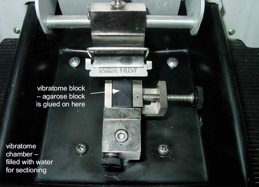
- Glue trimmed agarose block onto black vibrating microtome block with superglue (cyanoacrylate).Trim block carefully so that top and bottom are parallel before glueing down.Line up on vibrating microtome block first to check before glueing.
- Fill vibrating microtome chamber with water or buffer until it comes up to the top of the chuck.Place vibrating microtome block with sample into chuck and clamp firmly.
- Insert half of double-edged razor blade, or injection blade, into holder.Set blade angle to between 10° and 25° in the first instance.
- Wind chuck up or down so that the top of the agarose block is just below the blade.Top up vibrating microtome chamber so water/buffer level just touches the blade and covers the specimen.
- Set amplitude and speed at about the middle to start with.Advance the blade, check that it does not begin to slice off a large chunk of tissue.
- Advance the blade until it begins to section through the agarose and tissue.Use section thickness of 50-200 μm, depending on cell size.
- Collect each section carefully with forceps and place on a sticky slide (coated with silane, polyethyleneimine, poly-L-lysine or other adhesive material).Keep slides in a moist chamber until sectioning is finished.
- Stain the sections! (staining protocol yet to be added)

LITERATURE REFERENCES
RL Peterson, CA Peterson and LH Melville (2008). ‘Teaching Plant Anatomy’. NRC Press, Ottawa, Canada ISBN 978-0-660-19798-2:This book is all about hand sectioning a wide range of tissues and observing either unstained or stained and it has a CD with it.
HEALTH, SAFETY & HAZARDOUS WASTE DISPOSAL CONSIDERATIONS
Be extremely careful with microtome blades!
If you leave the microtome unattended and a blade is mounted then place a sign over the blade to reduce risk of cuts.